
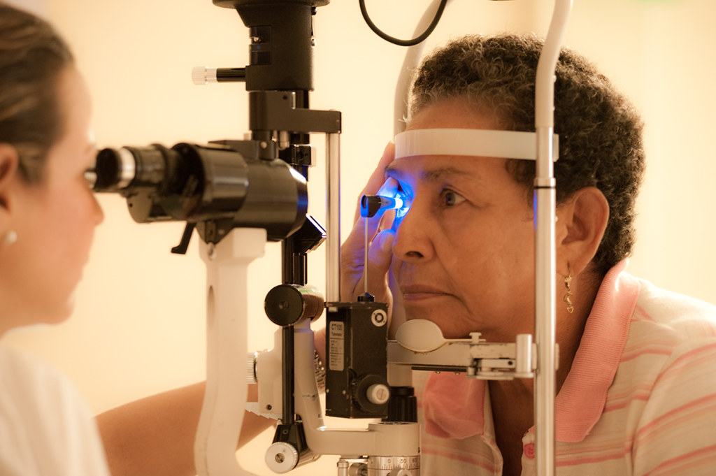
Diagnostic imaging plays a crucial role in understanding eye health. It helps us catch and treat eye conditions early. From simple scans to detailed images, each technique provides valuable insights. Technologies like OCT, ultrasound, and fundus photography give us a clear view of the eye’s structure. Invision Sally Jobe is one pioneer using advanced imaging in eye care. This exploration will shine a light on how these tools improve diagnosis and treatment.
Understanding OCT: Optical Coherence Tomography
Optical Coherence Tomography (OCT) is a non-invasive imaging test. It utilizes light waves to take cross-section pictures of your retina. With OCT, we can see each of the retina’s distinctive layers. This allows us to map and measure their thickness. These measurements help in the early detection and diagnosis of retinal diseases like glaucoma and age-related macular degeneration (AMD). More information on OCT can be found at the National Eye Institute.
Ultrasound in Eye Care
Ultrasound uses sound waves to create images of the eye. It is especially useful for examining the inside of the eye when the view is blocked due to cataracts or other conditions. This method helps in diagnosing and monitoring tumors, retinal detachments, and other serious eye conditions. The National Institute of Biomedical Imaging and Bioengineering provides further details on ultrasound imaging.
Fundus Photography: A Picture of Eye Health
Fundus photography involves capturing detailed images of the interior surface of the eye. This includes the retina, optic disc, macula, and posterior pole. These images are crucial for monitoring the progression of eye diseases and assessing the health of the eye. By comparing photographs taken over time, we can track changes and make informed treatment decisions.
Comparing Imaging Techniques
Different imaging techniques serve different purposes. Here’s how they compare:
| Imaging Technique | Best For | Limitations |
| OCT | Retinal thickness and layers | Limited in patients with media opacities |
| Ultrasound | Internal eye conditions with obstructions | Lower resolution compared to OCT |
| Fundus Photography | Monitoring disease progression | Does not show retinal layers |
Advancements and Future Directions
The field of diagnostic imaging is constantly evolving. New techniques and technologies are paving the way for better and more accurate diagnoses. Innovations such as adaptive optics and machine learning are starting to play a role in enhancing these imaging methods. These advancements could lead to faster and more personalized care, improving outcomes for patients.
The Importance of Early Detection
Early detection through imaging can prevent vision loss. Many eye diseases, if caught early, can be managed effectively. Diagnostic imaging allows doctors to detect issues before symptoms arise. This proactive approach is vital in preserving eyesight and maintaining quality of life.
Conclusion
Diagnostic imaging in ophthalmology is a powerful tool in eye care. Techniques like OCT, ultrasound, and fundus photography provide essential insights into eye health. These technologies enhance our ability to diagnose and treat eye conditions with precision. By staying informed and embracing these advancements, we ensure the best possible care for our eyes.